Image 18635: Anatomy of the Spine Illustration
Description
Description
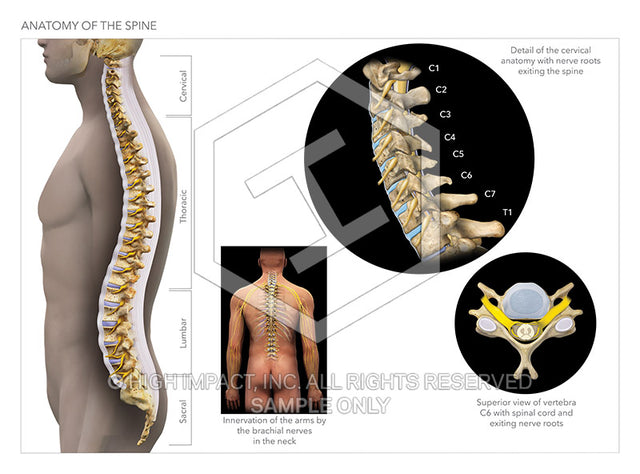
This graphic includes four illustrations of normal spinal anatomy focusing on the cervical spine. The first is a lateral or sagittal view of the entire spine showing normal vertebra, disc and peripheral nerve anatomy including labels for the cervical, thoracic, lumbar and sacral areas. The second illustration focuses on the peripheral nerves of the cervical and thoracic regions. The third illustration is a lateral or side view of the cervical spine featuring the vertebra, discs and nerve roots. The fourth and final image is an axial view of a cervical vertebra with associated intervertebral disc, facet joints, spinal cord, and nerve roots. Best used to demonstrate normal cervical anatomy in comparison to traumatic spinal injuries demonstrated in a separate graphic showing the abnormalities post-trauma.
Best used for demand letters or narrative reports in traumatic cervical spinal injury cases as well as for use in mediation, arbitration or trial for traumatic spinal injuries as an example of the anatomy before injury.
To purchase a single-user perpetual license to this stock image, you must first agree to the terms of service with High Impact and Trial Guides. Please note: the use of this image in any marketing is strictly prohibited and is a violation of the terms. Your agreement to the terms will apply to all future High Impact image purchases. Upon the completion of your purchase, an email will be sent to you containing a download link for a high-resolution TIFF file ready for your immediate use. No physical product will be delivered.
For customization requests, you can contact High Impact directly at www.highimpact.com.